
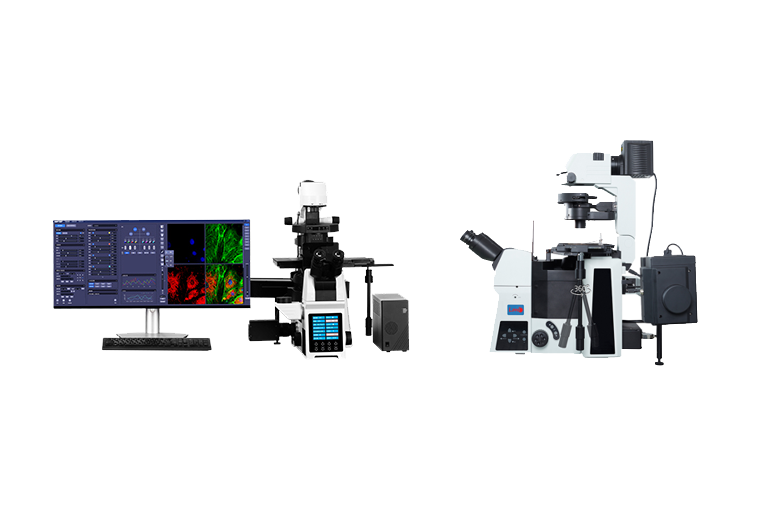
Life science
Cells, tissues & microorganisms
Bright-field, phase-contrast, and fluorescence approaches support observation across biological specimens.
Discuss this applicationDifferent specimens and workflows call for different microscopy approaches. Explore common application areas, then contact LMI for current system options.
Explore applications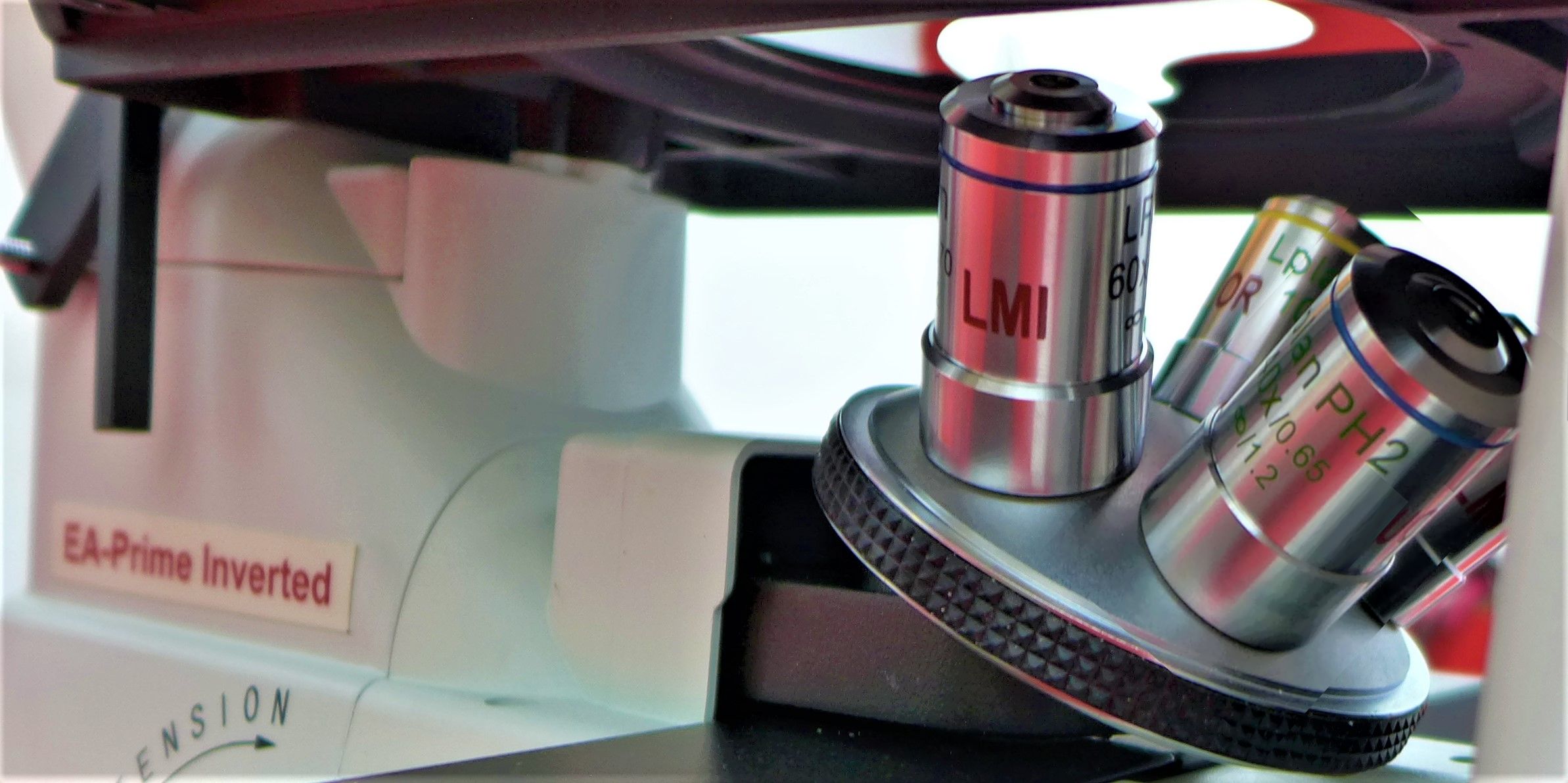
The categories below describe common uses of optical and digital microscopy without limiting the system configuration.
Bright-field, phase-contrast, and fluorescence approaches support observation across biological specimens.
Discuss this application
Microscopy helps learners understand specimen preparation, focusing, illumination, and structured observation.
Discuss this application
Digital imaging enables live display, image capture, sharing, annotation, and record keeping.
Discuss this application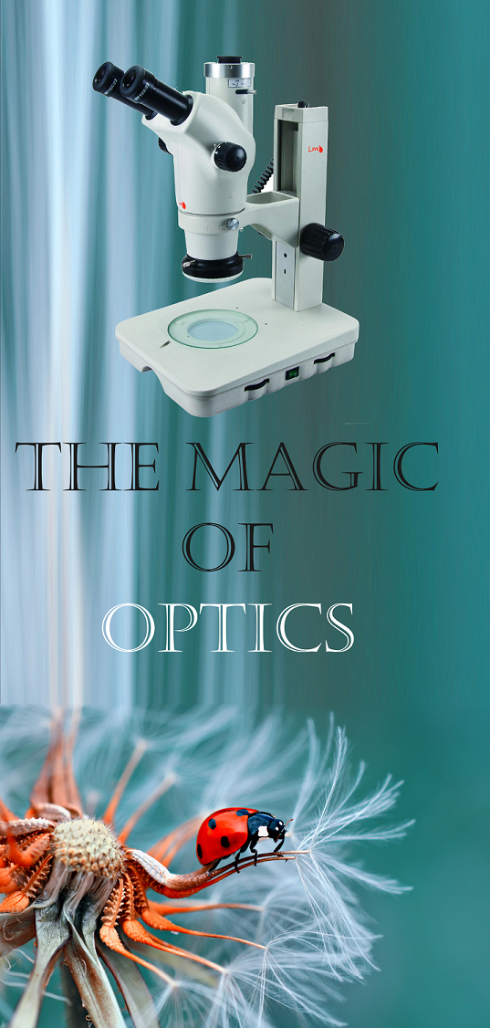
Three-dimensional, low-magnification viewing supports manipulation and examination of larger specimens.
Discuss this applicationOptical and digital microscopy can support surface viewing, component inspection, and visual documentation.
Discuss this applicationA suitable configuration depends on specimen type, contrast requirements, viewing time, and output needs.
Discuss this applicationSearch by industry, specimen, process, or inspection requirement. Open any section to view its typical microscopy uses.
Showing 28 application areas
| Microscope type | Typical applications |
|---|---|
| Biological | Biology, medicine, microbiology, and education |
| Metallurgical | Metals, alloys, welding, and failure analysis |
| Polarizing | Minerals, crystals, geology, and polymers |
| Stereo | Electronics, assembly, surgery, entomology, and inspection |
| Fluorescence | Cell biology, immunology, and molecular biology |
| Digital | Documentation, teaching, quality control, and remote collaboration |
| Industrial | Manufacturing, precision engineering, and surface inspection |
| Inverted | Cell culture, live-cell imaging, and IVF laboratories |
| Comparison | Forensic investigations |
| Phase contrast | Live, unstained cells and microorganisms |
| Darkfield | Fine particles, microorganisms, and transparent specimens |
These general questions help define the optical and imaging requirements before choosing a system.
Bright-field observation is commonly associated with specimens that already provide sufficient contrast, including many stained preparations.
Phase-contrast techniques can increase the visibility of transparent specimens without requiring conventional staining.
Digital imaging requirements may include live viewing, capture resolution, documentation, presentation, or measurement.
Stereo zoom microscopy provides depth perception and working space for dissection, positioning, assembly, or inspection tasks.
Share the specimen type, smallest detail, contrast method, and required output with the LMI team.