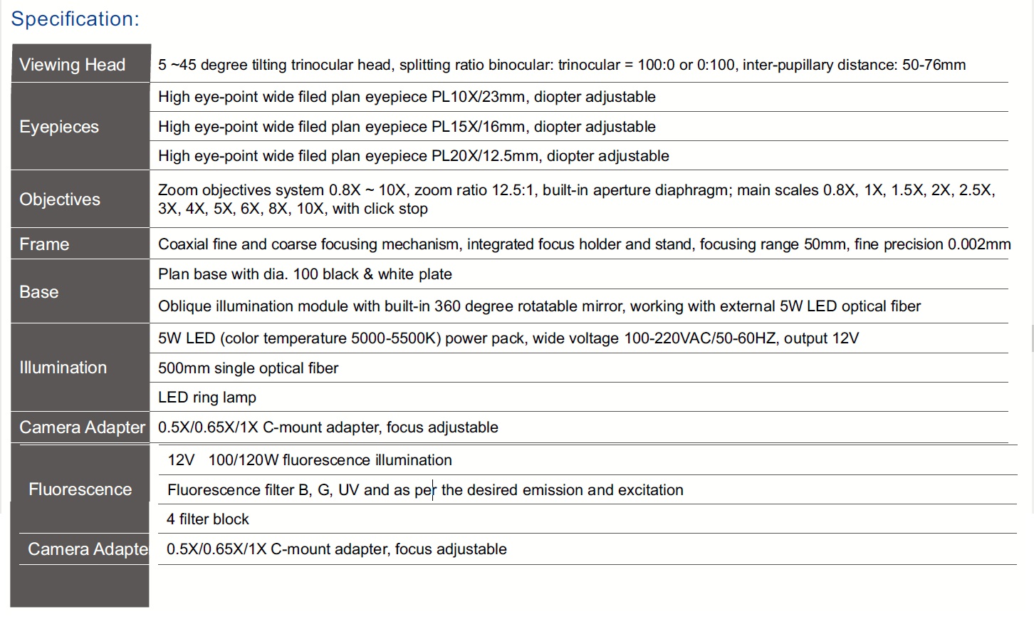

High-end research platform
A sophisticated and comfortable stereo imaging system for life science, microelectronics and semiconductor applications.
High-end research stereo microscope
A Galileo stereo zoom platform engineered for life science, microelectronics and semiconductor research, combining a large zoom range with precise imaging and ergonomic control.
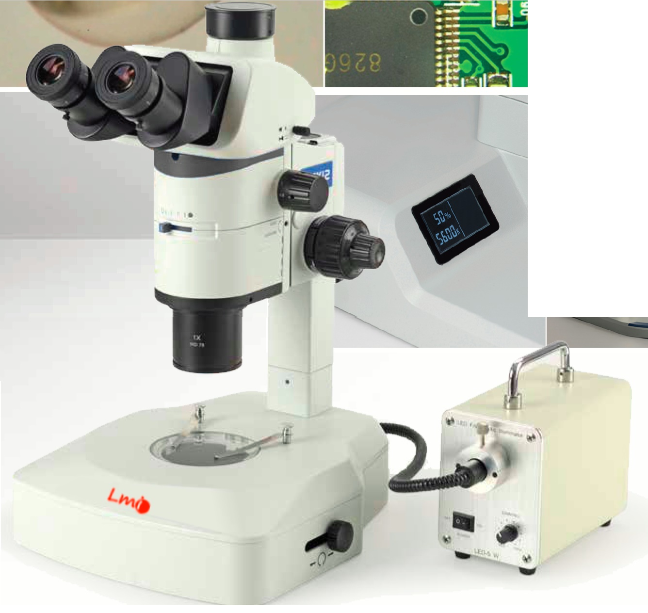
Research without compromise
The SZM-12 supports biological experiments, chemical analysis, cell culture, microelectronics and semiconductor inspection. Its apochromatic design corrects axial chromatic aberration across red, green, blue and violet wavelengths, converging them on one focal plane for faithful sample colour.
Ergonomic controls and a flexible viewing head reduce fatigue during extended observation, while selectable click stops provide repeatable magnification or smooth continuous zooming.


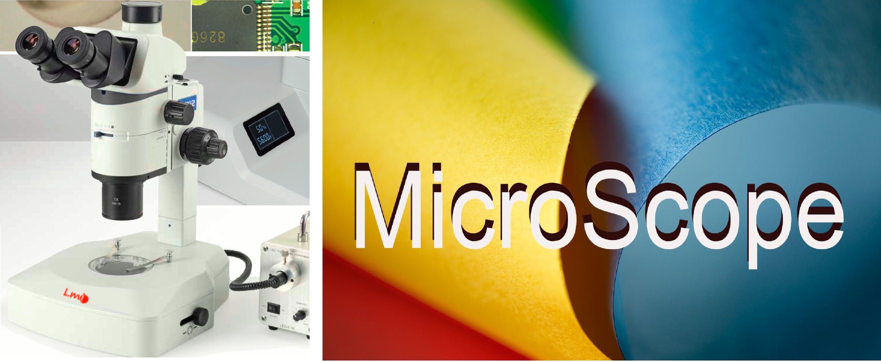
System highlights
A sophisticated and comfortable stereo imaging system for life science, microelectronics and semiconductor applications.

Advanced correction brings red, green, blue and violet wavelengths into a shared focal plane, improving colour rendition and presenting samples with natural, accurate colour.
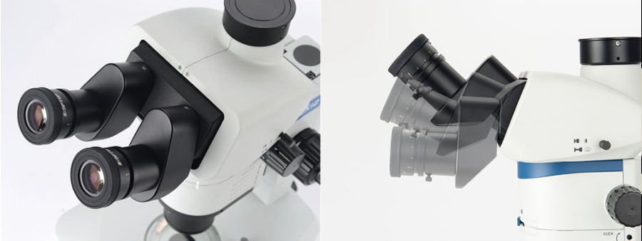
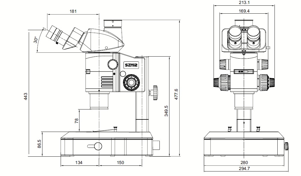
The viewing angle adjusts from 5° to 45°, adapting to different operators and working postures for more comfortable extended observation.
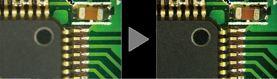
A front-mounted aperture adjustment provides direct control over depth of field for higher-quality images across varied samples.
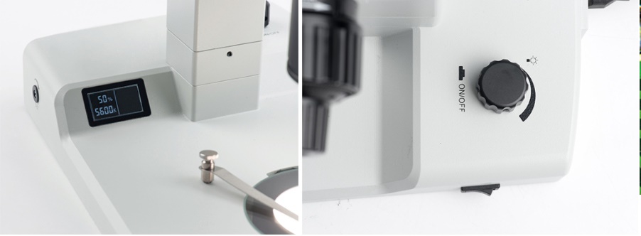
Adjust light intensity with the dimmer and read the exact brightness percentage on the base LCD, making it easier to record and reproduce the best illumination for each sample.
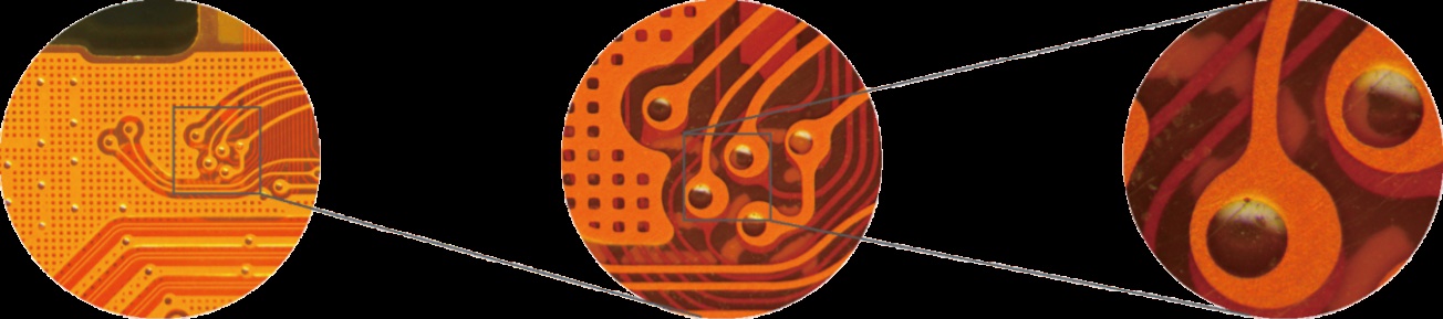
Zoom continuously from 0.8X to 10X. Main magnifications have click stops for repeatability, which can be disengaged whenever smooth continuous zoom is preferred.

The man-machine engineered Galileo system produces detailed three-dimensional images with intuitive operation for biomedical and industrial research.
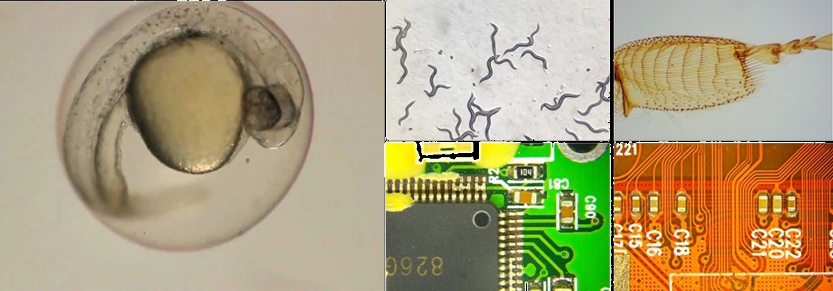
Stress-free cold-light illumination delivers even, reflection-free fields across the zoom range. Optional fluorescence provides efficient detection of weak signals, while a large object field, working distance and depth of field simplify specimen manipulation.
A separate camera-focusing mechanism aligns the live screen image with the eyepiece focus. Once set, users can capture images while retaining maximum optical precision through the eyepieces.